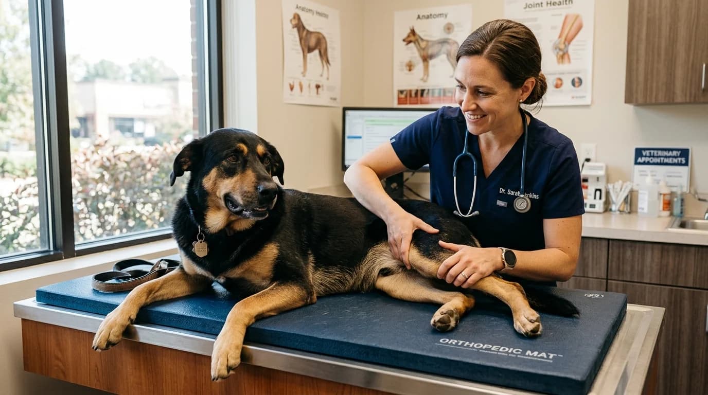
Canine Osteoarthritis (OA)—also recognized clinically as Degenerative Joint Disease (DJD)—represents the single most prevalent chronic debilitating condition encountered in small animal veterinary medicine. Epidemiological investigations establish that over 20% of all dogs older than one year and more than 80% of canines over eight years exhibit radiographic and histopathological evidence of joint degeneration.
In large and giant breeds—such as German Shepherds, Labrador Retrievers, Golden Retrievers, Rottweilers, Bernese Mountain Dogs, and Great Danes—this vulnerability is intensified exponentially. Biomechanical load scaling means that somatic weight amplifies joint stress, accelerating the degradation of articular cartilage, subchondral bone remodeling, and chronic low-grade synovial inflammation.
For decades, veterinary care treated canine arthritis reactively: waiting until an older dog presented with overt lameness, morning stiffness, or refusal to ascend stairs before prescribing non-steroidal anti-inflammatory drugs (NSAIDs). Today, the paradigm has shifted decisively toward proactive, multimodal joint preservation.
By leveraging early phenotypic screening (such as the PennHIP distraction index at 16 weeks), monitoring emerging serum and synovial molecular biomarkers, implementing Disease-Modifying Osteoarthritis Drugs (DMOADs), administering targeted neuro-modulating biologics, and re-engineering home microenvironments, veterinary clinicians and dedicated pet parents can arrest cartilage catabolism years before visible clinical disability takes hold.
Pathophysiology of Canine OA: The Molecular Cascade
Osteoarthritis is far more than mechanical "wear-and-tear." It is an active, metabolically driven inflammatory disease affecting the entire diarthrodial organ—encompassing the articular cartilage, subchondral bone plate, synovium, fibrous joint capsule, menisci, and periarticular soft tissues.
+-------------------------------------------------------------------------+
|                PRIMARY BIOMECHANICAL INSULT OR DYSPLASIA                |
|      (Joint Incongruity, Synovial Laxity, Repetitive Micro-Trauma)      |
+-------------------------------------------------------------------------+
                                     |
                                     v
+-------------------------------------------------------------------------+
|                  CELLULAR CHONDROCYTE PHENOTYPIC SHIFT                  |
|    Upregulation of IL-1beta & TNF-alpha; Degradative Protease Secretion  |
+-------------------------------------------------------------------------+
                                     |
                                     v
+-------------------------------------------------------------------------+
|                EXTRACELLULAR MATRIX (ECM) CATABOLISM                    |
|   • MMP-13 Cleaves Type II Collagen Fibers (Irreversible Loss)          |
|   • ADAMTS-4 & ADAMTS-5 Degrade Aggrecan Proteoglycans                  |
|   • Loss of Chondroitin Sulfate -> Loss of Bound Articular Water        |
+-------------------------------------------------------------------------+
                                     |
                                     v
+-------------------------------------------------------------------------+
|                 SYNOVIAL & SUBCHONDRAL SCLEROSIS CYCLE                  |
|   Synovitis -> VEGF & Nerve Growth Factor (NGF) Sprouting -> Osteophytes|
|                  Peripheral & Central Sensitization (Pain)              |
+-------------------------------------------------------------------------+
1. The Chondrocyte and the Extracellular Matrix (ECM)
Normal articular cartilage is an avascular, aneural tissue populated by specialized cells called chondrocytes. These cells maintain an intricate Extracellular Matrix composed primarily of:
- Type II Collagen Fibrils (90%–95% of total collagen): Forms a tensile, cross-linked structural scaffolding that holds the tissue together under shear forces.
- Aggrecan: Large proteoglycan monomers decorated with hydrophilic glycosaminoglycan (GAG) side chains—specifically chondroitin sulfate and keratan sulfate—attached to a hyaluronic acid backbone. Aggrecan attracts water molecules under osmotic pressure, creating hydrostatic fluid pressurization that cushions compressive loads.
2. Pro-Inflammatory Cytokines & Matrix Metalloproteinases
When mechanical instability (such as hip or elbow dysplasia) exerts shear stress on articular surfaces, chondrocytes undergo an aberrant phenotypic shift. They release pro-inflammatory cytokines, predominantly Interleukin-1 beta (IL-1$\beta$) and Tumor Necrosis Factor-alpha (TNF-$\alpha$).
These cytokines trigger gene transcription for aggressive catabolic enzymes:
- Aggrecanases (ADAMTS-4 and ADAMTS-5): Sever the interglobular domain of aggrecan core proteins, causing immediate depletion of water-binding proteoglycans.
- Matrix Metalloproteinase-13 (MMP-13 / Collagenase-3): Specifically targets and cleaves the triple helix of type II collagen. Because adult articular chondrocytes possess virtually zero capacity to synthesize replacement type II collagen, this cleavage represents an irreversible structural breakdown.
3. Subchondral Bone Remodeling & Osteophytosis
As the protective cartilage surface thins and fibrillation occurs, mechanical force transfers directly onto the underlying subchondral bone. The bone undergoes micro-fractures, followed by dense, sclerotic thickening (eburnation). At the non-weight-bearing joint margins, mechanical strain and vascular endothelial growth factor (VEGF) promote endochondral ossification, generating sharp, bony spurs known as osteophytes and enthesophytes.
4. Synovitis and Pain Neurobiology: Peripheral to Central Sensitization
Fibrillated cartilage micro-fragments shed directly into the joint lumen, phagocytosed by type A synoviocytes. This chronic foreign-body challenge incites synovitis, flooding the joint fluid with prostaglandins ($PGE_2$), substance P, bradykinin, and Nerve Growth Factor (NGF).
NGF binds to tropomyosin receptor kinase A (TrkA) receptors on sensory nociceptive fibers, causing peripheral sensitization (hyperalgesia at the joint). Left untreated, continuous nociceptive bombardment into dorsal root ganglia stimulates spinal NMDA receptors, cementing central sensitization ("wind-up" pain), where even non-noxious movements trigger excruciating discomfort.
Large Breed Predisposition & Anatomical Vulnerability
Due to rapid somatotropic growth trajectories, elevated body mass, and selective breeding architecture, distinct large and giant breeds exhibit well-characterized anatomical vulnerabilities:
| Breed | Weight Class (Adult) | Primary Anatomical Joint Vulnerability | Primary Underlying Etiology | Recommended Initial Screening Age |
|---|---|---|---|---|
| German Shepherd | 65–90 lbs (29–41 kg) | Coxofemoral (Hip) & Lumbosacral (L7–S1) | Joint laxity, hip dysplasia, lumbosacral stenosis | 16 wks (PennHIP), 6–12 mos (Spine) |
| Labrador Retriever | 55–80 lbs (25–36 kg) | Cubital (Elbow) & Stifle (Knee) | Fragmented Coronoid Process (FCP), Cranial Cruciate Ligament (CCL) rupture | 16–24 wks (Elbow CT / PennHIP) |
| Golden Retriever | 55–75 lbs (25–34 kg) | Coxofemoral (Hip) & Cubital (Elbow) | Ununited Anconeal Process (UAP), hip laxity, early ligamentous strain | 16 wks (PennHIP / Elbow Rads) |
| Rottweiler | 80–135 lbs (36–61 kg) | Cubital (Elbow) & Stifle (CCL) | Medial Coronoid Disease (MCD), bilateral cruciate instability | 16–20 wks (CT arthrography) |
| Bernese Mountain Dog | 70–115 lbs (32–52 kg) | Cubital (Elbow) & Coxofemoral | Elbow incongruity, severe polyarticular developmental dysplasia | 16 wks (PennHIP & Multi-view Elbow) |
| Great Dane | 110–180+ lbs (50–82+ kg) | Stifle, Carpal, & Cervical Vertebrae | Rapid osteochondrosis dissecans (OCD), Wobbler syndrome | 16–24 wks (Radiographic screening) |
| Newfoundland | 100–150 lbs (45–68 kg) | Stifle (CCL) & Coxofemoral | Ligamentous hyperlaxity, deep coxofemoral subluxation | 16 wks (PennHIP) |
| Cane Corso / Mastiff | 90–140+ lbs (41–64+ kg) | Coxofemoral, Stifle, & Tarsus (Hock) | Massive axial compressive load, tibial plateau angle (TPA) deformity | 16–20 wks (Orthopedic evaluation) |
To determine your dog's exact biological age stage and compare aging velocity across weight classifications, explore our interactive Dog Age Calculator or consult the Dog Age Chart.
Early Biomarkers & Objective Diagnostics
The traditional gold standard of diagnosing canine OA via plain orthogonal radiography has an inherent limitation: radiographs only detect structural osseous changes after significant, irreversible cartilage damage has already transpired.
Modern veterinary orthopedics utilizes proactive clinical assessment tools capable of identifying joint vulnerability in the juvenile and early adult stages:
                            DIAGNOSTIC TIME HORIZON
  Juvenile (4-6 Mos)             Young Adult (1-3 Yrs)           Mature / Senior (4+ Yrs)
+-----------------------+     +------------------------+     +--------------------------+
| PennHIP Screening     |     | Synovial Biomarkers    |     | Plain Radiography (OFA)  |
| • Distraction Index   | --> | • CTX-II & COMP        | --> | • Subchondral Sclerosis  |
| • Joint Laxity Risk   |     | • Hyaluronic Acid      |     | • Periarticular Spurs    |
| • JPS Candidacy       |     | Force Plate Kinematics |     | • Joint Space Collapse   |
+-----------------------+     +------------------------+     +--------------------------+
1. PennHIP Distraction Index (DI) at 16 Weeks
The University of Pennsylvania Hip Improvement Program (PennHIP) is a stress-radiographic technique performed under deep sedation or general anesthesia as early as 16 weeks of age.
- Unlike standard hip-extended ventrodorsal views (which mechanically wind the joint capsule tight, artificially concealing passive laxity), PennHIP measures the Distraction Index (DI)—a quantitative ratio of femoral head displacement relative to the acetabulum under a standardized distractor force.
- DI Interpretation:
- $\text{DI} < 0.30$: Tight hips; virtually zero risk of developing coxofemoral osteoarthritis throughout life.
- $\text{DI} = 0.30 \text{ to } 0.69$: Moderate laxity; variable risk requiring active environmental and nutritional intervention.
- $\text{DI} \ge 0.70$: Severe joint laxity; near 100% probability of secondary osteoarthritic degradation without surgical intervention.
- Juvenile Pubic Symphysiodesis (JPS) Window: If severe laxity ($\text{DI} > 0.5$) is identified before 16 to 20 weeks of age, minimally invasive thermal cauterization of the pubic symphysis (JPS) alters subsequent pelvic growth, rotating the acetabular rims laterally over the femoral heads to permanently stabilize the joints.
2. Molecular Serum and Synovial Fluid Biomarkers
Veterinary research has validated multiple molecular fragments shed into biological fluids during cartilage breakdown:
- CTX-II (C-terminal telopeptide of type II collagen): A direct degradation fragment created when MMP-13 cleaves type II collagen fibrils. Elevated urinary or serum CTX-II concentrations serve as a quantitative biomarker of ongoing active collagen loss.
- COMP (Cartilage Oligomeric Matrix Protein): A non-collagenous structural glycoprotein involved in fibril assembly. Spikes in synovial COMP reflect accelerated extracellular turnover.
- Synovial Hyaluronic Acid (HA) Depolymerization: Healthy synovial fluid has high viscoelasticity driven by high-molecular-weight HA ($> 2.5 \times 10^6\text{ Da}$). During early synovitis, reactive oxygen species depolymerize HA into low-molecular-weight fragments ($< 0.5 \times 10^6\text{ Da}$), drastically reducing fluid boundary lubrication.
3. Objective Kinetic Gait Analysis (Force Plates & Pressure Mats)
Canine patients are notorious for concealing subtle bilateral pain. Visual lameness scoring by veterinarians or owners suffers from heavy subjective bias (placebo caregiver effect).
- Ground Reaction Forces (GRF): Measuring Peak Vertical Force (PVF) and Vertical Impulse (VI) via computerized piezoelectric force platforms detects weight-bearing asymmetries as minor as 2% to 3%, enabling clinical intervention months before visible head-bobs or limps emerge.
Canine Osteoarthritis Staging: The COAST System
Developed by an international consensus of veterinary orthopedic surgeons, the Canine Osteoarthritis Staging Tool (COAST) establishes a standardized framework for grading joint disease and guiding stage-matched therapies:
| COAST Stage | Clinical Definition | Patient Demeanor & Activity | Radiographic Findings | Primary Clinical Objective |
|---|---|---|---|---|
| Stage 0 (Pre-Clinical) | Dog is clinically sound but has genetic or anatomical risk factors (e.g., German Shepherd puppy, high-drive sporting dog). | 100% normal energy, enthusiasm, and symmetric weight-bearing. | No radiographic changes; zero osteophytes. | Primary prevention: PennHIP screening, controlled growth nutrition, avoid concussive trauma. |
| Stage 1 (Subclinical) | Joint laxity or early structural incongruity present; no clinical signs observed by owner. | Normal gait; subtle reluctance to leap into high vehicles or slight hesitation after hard exercise. | Trace osteophytosis ($< 1\text{ mm}$); minimal sclerosis; elevated distraction index ($\text{DI} > 0.4$). | Secondary prevention: Initiate DMOADs (Adequan), therapeutic marine EPA/DHA, core conditioning. |
| Stage 2 (Mild OA) | Mild lameness; stiffness after rest ("warming out" of stiffness within 5 minutes). | Mild exercise intolerance; subtle shifts in weight distribution while eating or standing. | Definite osteophytes ($1\text{ to } 2\text{ mm}$); mild subchondral sclerosis; altered synovial fluid viscosity. | Active multimodal management: Oral chondroprotectants (UC-II), PRN NSAID / Galliprant, low-impact conditioning. |
| Stage 3 (Moderate OA) | Obvious persistent or intermittent lameness; pain on full joint extension/flexion during exam. | Frequent stiffness; bunny-hopping upstairs; difficulty rising from hard surfaces; audible joint crepitus. | Moderate-to-severe osteophytes ($3\text{ to } 5\text{ mm}$); joint remodeling; loss of diarthrodial space. | Comprehensive medical therapy: Librela (anti-NGF mAb) or chronic NSAIDs, physical hydrotherapy, home ergonomics. |
| Stage 4 (Severe OA) | Severe chronic lameness; marked functional disability; significant muscular sarcopenia. | Reluctance to walk; groaning when lying down; constant postural compensation; behavioral irritability. | Complete collapse of joint space; massive osteophytic bridging; severe bone eburnation; periarticular fibrosis. | Advanced palliative / surgical care: Multimodal multimodal analgesia (gabapentin, amantadine), joint injections, salvage arthroplasty (THR). |
Pharmacological & Biologic Landscape: DMOADs, Anti-NGF mAbs & NSAIDs
Effective long-term OA management in large breeds avoids sole reliance on a single pill. Instead, it coordinates disease-modifying agents that protect remaining cartilage with precise analgesic therapies that disrupt pain signaling.
| Class | Medication | Mechanism of Action | Administration Route | Dosing Frequency | Primary Target Stage | Clinical Monitoring & Considerations |
|---|---|---|---|---|---|---|
| DMOAD | Adequan Canine (Polysulfated Glycosaminoglycan - PSGAG) | Inhibits MMPs and aggrecanases; stimulates endogenous HA synthesis; chondroprotective | Intramuscular (IM) injection | Loading: 4.4 mg/kg twice weekly for 4 wks; maintenance every 2–4 wks | COAST Stages 1–3 | Monitor coagulation if bleeding disorders present; gold standard for early structural preservation. |
| DMOAD | Cartrophen Vet (Pentosan Polysulfate Sodium - PPS) | Stimulates hyaluronan synthesis; enhances microvascular subchondral perfusion; inhibits catabolism | Subcutaneous (SC) injection | 3 mg/kg once weekly for 4 consecutive weeks; repeat cycle every 3–6 mos | COAST Stages 1–3 | Mild transient anticoagulant activity; excellent cartilage matrix stabilizer. |
| Anti-NGF mAb | Librela (Bedinvetmab) | Fully caninized IgG monoclonal antibody targeting Nerve Growth Factor (NGF); blocks pain signaling | Subcutaneous (SC) injection | 0.5 – 1.0 mg/kg once every 28 days | COAST Stages 2–4 | Does not target cartilage directly (pure analgesic); do not use in breeding/pregnant animals; monitor for rapid progression if joint unstable. |
| EP4 Receptor Antagonist | Galliprant (Grapiprant) | Non-COX inhibiting prostaglandin receptor antagonist; selectively blocks EP4-mediated OA pain | Oral tablet | 2 mg/kg once daily | COAST Stages 2–3 | Spares COX-1 and COX-2; significantly lower risk of GI ulceration and renal hypoperfusion than standard NSAIDs. |
| COX-2 Inhibiting NSAID | Carprofen / Meloxicam / Firocoxib | Selective inhibition of cyclooxygenase-2 (COX-2); blocks downstream synthesis of inflammatory $PGE_2$ | Oral tablet or liquid | Carprofen: 2.2 mg/kg BID or 4.4 mg/kg SID; Meloxicam: 0.1 mg/kg SID | COAST Stages 2–4 | Baseline and biannual serum biochemistry (ALT, ALKP, BUN, Creatinine) mandatory; high efficacy for active flares. |
| NMDA Antagonist | Amantadine | Blocks spinal cord N-methyl-D-aspartate (NMDA) receptors; reverses central sensitization & "wind-up" | Oral capsule | 3 – 5 mg/kg once daily (combined with NSAID/mAb) | COAST Stages 3–4 | Excellent adjunct for chronic refractory central pain; minimal organ toxicity. |
1. Disease-Modifying Osteoarthritis Drugs (DMOADs)
True DMOADs represent the pinnacle of early proactive medical intervention. Unlike NSAIDs, which only relieve pain symptoms downstream, DMOADs (such as Adequan Canine and Pentosan Polysulfate) physically incorporate into the articular cartilage matrix:
- Mechanism: Inhibit serine proteinases, elastases, and MMP-13; suppress complement activation; stimulate synovial synoviocytes to synthesize high-molecular-weight endogenous hyaluronic acid.
- Optimal Timing: Initiating Adequan at COAST Stage 1 or 2—rather than waiting for Stage 4 terminal sclerosis—delays macroscopic joint deterioration by years.
2. The Biologic Revolution: Anti-NGF Monoclonal Antibodies (Bedinvetmab / Librela)
Approved globally as the first caninized monoclonal antibody for canine OA pain, Bedinvetmab (Librela) has transformed senior dog management:
- Mode of Action: Neutralizes Nerve Growth Factor (NGF) in interstitial fluid, preventing it from binding to TrkA receptors on peripheral nociceptors and desensitizing local afferent pain fibers.
- Metabolism: Being a therapeutic antibody, bedinvetmab is degraded into constituent peptides and amino acids via standard reticuloendothelial catabolism, completely bypassing hepatic and renal cytochrome P450 pathways. This makes it uniquely advantageous in geriatric patients with subclinical renal or liver elevations.
- Clinical Nuance: Because Librela is an extraordinary analgesic, dogs frequently experience a sudden surge in energy and mobility. Guardians must ensure high-energy large dogs do not engage in explosive ball chasing, which could physically overload mechanically unstable joints.
Nutritional Architecture & Evidence-Based Nutraceuticals
Nutritional management forms the biological foundation of joint cartilage integrity. In large breeds, this strategy is dual-pronged: metabolic body weight optimization and targeted synovial chondroprotection.
                           CANINE CHONDROPROTECTIVE TRIAD
                                      [ EPA / DHA ]
                              (Competitive Arachidonic Blockade;
                                100-150 mg/kg Daily Dose)
                                      /          \
                                     /            \
                                    v              v
                  [ UC-II COLLAGEN ]  <--------->  [ PERNA CANALICULUS ]
                 (Oral GALT Tolerance;               (ETA, Glycosaminoglycans,
                  Deactivates T-Cell Attack)          Natural Anti-Inflammatory)
1. The Adipose-Inflammation Axis & Caloric Control
Adipose tissue is not an inert storage depot; it is an active endocrine organ that secretes adipokines (leptin, adiponectin, resistin) and pro-inflammatory cytokines (TNF-$\alpha$, IL-6). In large dogs, obesity exerts a destructive double penalty:
- Excess Static Load: Amplifies compressive stress on load-bearing stifle and hip surfaces.
- Systemic Inflammaging: Circulating adipokines accelerate baseline synovial cartilage catabolism.
- Target Body Condition: Maintain large breeds at a lean Body Condition Score (BCS) of 4 to 4.5 out of 9 (palpable ribs with minimal fat cover, distinct waistline when viewed from above). Landmark lifelong Purina canine studies proved that maintaining a lean body condition delayed radiographic osteoarthritis onset by an average of nearly two full years.
2. Therapeutic Marine Omega-3 Fatty Acids (EPA & DHA)
Dietary long-chain omega-3 polyunsaturated fatty acids displace arachidonic acid (omega-6) in chondrocyte membrane phospholipids:
- Target Dosage: 100 to 150 mg combined EPA + DHA per kg of body weight daily.
- Clinical Effect: Suppresses leukotriene $B_4$ and $PGE_2$ production, downregulating MMP-13 gene expression within 60 to 90 days of consistent administration.
- Note: Plant-based flaxseed oil (ALA) is ineffective in dogs due to near-zero enzymatic conversion by canine delta-6-desaturase. Purified cold-water marine fish oils or concentrated algae oils are clinically required.
3. Undenatured Type II Collagen (UC-II)
Unlike hydrolyzed collagen (which acts purely as basic amino acid building blocks), Undenatured Type II Collagen (UC-II) retains its native tertiary triple-helix epitope structure:
- Mechanism (Oral Tolerization): As intact UC-II passes through the small intestine, it interacts with gut-associated lymphoid tissue (GALT / Peyer's patches). This trains naive T cells into regulatory T-cells ($T_{\text{reg}}$) specific for type II collagen epitopes.
- When these $T_{\text{reg}}$ cells migrate through circulation to arthritic joints, they recognize endogenous collagen fibrils and release anti-inflammatory cytokines (IL-10, TGF-$\beta$), halting autoimmune-mediated joint destruction. Controlled clinical trials demonstrate superior pain reduction in dogs compared to traditional glucosamine/chondroitin combinations.
4. Green-Lipped Mussel (Perna canaliculus)
A marine bivalve indigenous to New Zealand, rich in unique omega-3 eicosatetraenoic acids (ETA), complex glycosaminoglycans, and bioactive zinc and manganese. ETA acts as a dual cyclooxygenase/lipoxygenase pathway inhibitor, alleviating periarticular stiffness.
For detailed guidance on tailoring nutrition, protein balance, and caloric density for mature canines, consult our comprehensive guide on Geriatric Canine Nutrition & Joint Support.
Home Ergonomics & Biomechanical Environmental Design
A comprehensive medical and nutritional protocol can be completely undermined if a large dog slips daily on slick hardwood floors or leaps out of a tall SUV. Designing an ergonomic home environment is a critical medical intervention:
1. Traction Infrastructure: Eliminating the "Splay" Trauma
Smooth flooring (hardwood, polished tile, laminate) presents an extreme biomechanical hazard for large dogs with early joint laxity or OA:
- Micro-Trauma Mechanism: Slipping triggers sudden, uncoordinated eccentric contractions of stabilizing adductor muscles and strains the cranial cruciate ligament and hip joint capsule.
- Ergonomic Remedy: Install non-skid runner rugs, interlocking foam mats, or heavy rubber-backed runners along primary indoor transit paths (hallways, around beds, eating areas).
- Direct Paw Solutions: Apply non-slip silicone traction grips (such as ToeGrips) or rubberized booties for dogs navigating mixed flooring.
2. Orthopedic Pressure Relieving Sleep Surfaces
A 90-pound dog lying on a thin pet bed compresses the cushion down to the hard floor, subjecting the greater trochanter of the femur, lateral humerus condyle, and lumbosacral spine to continuous focal pressure:
- Medical Bed Architecture: Invest in medical-grade multi-layer orthopedic beds featuring at least 4 to 6 inches of high-density base support foam bonded to high-resilience memory foam.
- Therapeutic Temperature: Provide beds with mild orthopedic contouring or low therapeutic thermal support; chronic arthritic joints stiffen when subjected to cold ambient drafts.
3. Ramp Incline vs. Jumping Forces
When a 70-pound Labrador leaps down from a vehicle tailgate or elevated furniture, the impact force transferred through the front carpal and elbow joints reaches 300% to 450% of the dog's static body weight:
- Vehicle & Furniture Ramps: Train large dogs early in adulthood to use textured, low-angle telescoping ramps ($\le 25^\circ\text{ incline}$) for vehicle entry and exit.
- Elevated Food & Water Bowls: Raised feeding stations aligned with the dog's mid-chest level prevent cervical flexion and anterior limb weight shifting during meals, reducing pressure on diseased elbows and the thoracic spine.
Physical Rehabilitation & Biomechanical Exercise Regimens
The physiological rule of thumb in canine joint care is: "Motion is lotion, but impact is injury." Cartilage has no blood vessels; it depends entirely on the cyclical compression and relaxation of joint movement to pump nutrient-rich synovial fluid through its matrix. Complete confinement induces cartilage thinning and rapid muscular sarcopenia. Conversely, explosive activities cause micro-fractures.
                          WEEKLY MOBILITY & CONDITIONING MATRIX
+-------------------+-------------------------------------------------------------+
| Exercise Modality | Clinical Protocol & Targeted Physiological Goal              |
+-------------------+-------------------------------------------------------------+
| Controlled Leash  | 15–20 mins, 2x daily on compliant turf/dirt; steady trotting |
| Walking           | maintains range of motion and prevents stiffness.           |
+-------------------+-------------------------------------------------------------+
| Hydrotherapy      | Underwater treadmill (UWTM) water at greater trochanter     |
| (UWTM)            | offloads 40–60% body weight, providing impact-free motion.  |
+-------------------+-------------------------------------------------------------+
| Targeted Proprio- | Cavaletti rails, balance wobble boards, weight-shifting     |
| ception Exercises | exercises rebuild stabilizing periarticular core muscles.   |
+-------------------+-------------------------------------------------------------+
| Passive Range of  | Gentle flexion/extension (10 reps/joint) combined with      |
| Motion (PROM)     | warm compresses to maintain capsular compliance.            |
+-------------------+-------------------------------------------------------------+
1. Controlled Low-Impact Aerobic Conditioning
- Structured Walk Strategy: Replace single long, fatiguing 45-minute hikes with two or three gentle 15- to 20-minute controlled walks on a standard 6-foot fixed leash.
- Surface Selection: Walk on shock-absorbing, compliant terrain (manicured turf, woodchip forest trails, packed sand) rather than concrete sidewalks or asphalt roadways.
- Eliminate Explosive Deceleration: Strictly eliminate high-intensity fetch games involving rapid sprint-stops, ball launchers, and erratic frisbee jumps, which deliver catastrophic shearing stress to the cranial cruciate ligaments.
2. Underwater Treadmill (UWTM) Hydrotherapy
The gold standard modality in veterinary physical rehabilitation:
- Buoyancy Mechanics: Filling the water basin to the level of the greater trochanter reduces static ground reaction forces by approximately 60%, allowing dogs with severe hip or stifle osteoarthritis to exercise vigorously without concussive impact.
- Warm Water Synovial Mobilization: Water heated to 86°F–90°F (30°C–32°C) stimulates vasodilation, relaxes tense myofascial trigger points, and softens periarticular capsular fibrosis.
3. Targeted Cavaletti Poles & Balance Training
- Cavaletti Rails: Stepping over spaced, low wooden poles (raised 2–4 inches off the floor) forces the dog to actively flex and extend the carpus, elbow, stifle, and hock through their full physiological range of motion rather than shuffling forward.
- Inflatable Balance Discs: Standing on compliant balance pads recruits deep stabilizing spinal multifidus and pelvic gluteal muscles, counteracting sarcopenia and stabilizing unstable joints.
Weekly Joint Health Management Checklist
Use this structured clinical management schedule to optimize joint preservation for your large or giant breed dog:
| Day | Primary Focus Area | Clinical Action Items | Primary Therapeutic Objective |
|---|---|---|---|
| Monday | Nutritional & Weight Audit | Weigh dog on veterinary scale or home pet scale; calculate exact daily caloric allotment; administer daily EPA/DHA & UC-II. | Prevents positive caloric drift; maintains lean Body Condition Score (BCS 4/9). |
| Tuesday | Low-Impact Turf Leash Walk | Two 20-minute steady-pace walks on soft grass or dirt trails; avoid abrupt stops. | Stimulates synovial fluid circulation; maintains baseline aerobic conditioning. |
| Wednesday | Home Ergonomics Inspection | Audit non-slip runner rug alignments; inspect orthopedic bed foam resilience; check ramp grip surface. | Eliminates slip hazards; ensures zero joint trauma from household mobility. |
| Thursday | Proprioception & Core Conditioning | 10 minutes of low cavaletti rail walking and gentle balance pad weight-shifting drills. | Re-educates proprioceptive nerve pathways; strengthens periarticular stabilizer muscles. |
| Friday | Controlled Walk & Passive Stretch | 15-minute gentle walk followed by 10 repetitions of gentle Passive Range of Motion (PROM) per limb. | Maintains full physiological capsular flexibility; mitigates weekend stiffness. |
| Saturday | Hydrotherapy or Gentle Sniffari | 20-minute supervised session in underwater treadmill (or gentle controlled swim in warm indoor canine pool). | Delivers maximum active joint mobilization with up to 60% weight offloaded. |
| Sunday | Rest, Myofascial Massage & Audit | 15 minutes of gentle myofascial trigger-point massage along lumbar and gluteal muscles; audit monthly DMOAD/Librela date. | Eases compensatory muscular strain; confirms adherence to medical injection schedules. |
Frequently Asked Questions
What are the earliest warning signs of arthritis in large dogs before they start limping?
The earliest signs of canine osteoarthritis are subtle behavioral and postural adjustments rather than dramatic limping:
- Hesitation Before Jumping: Brief pauses before hopping into a vehicle or onto elevated surfaces.
- Postural Weight Shifting: Standing with hind legs positioned unusually far forward under the belly, or shifting body weight forward onto the forelimbs while standing at the food bowl.
- Slowness Rising After Sleep: Taking 15 to 30 seconds to fully stand and stretch after lying down on a cold or hard surface.
- Behavioral Withdrawal or Irritability: Reluctance to greet owners enthusiastically at the doorway, or mild grumpiness when touched near the lower back or hips.
- Asymmetric Muscular Atrophy: Noticeable narrowing across the pelvic thigh musculature or widening of the shoulder girdle to compensate for rear-end discomfort.
How does early puppy growth speed affect arthritis risk later in life?
Rapid juvenile growth is a major contributing factor to developmental joint disease. Feeding high-calorie, high-protein puppy diets designed for small dogs accelerates longitudinal bone growth beyond the capacity of developing articular cartilage, leading to osteochondrosis dissecans (OCD) and joint incongruity. Large and giant breed puppies must consume diets formulated with controlled caloric density, moderate energy levels, and tightly balanced calcium-to-phosphorus ratios (1.1:1 to 1.3:1) to ensure slow, uniform skeletal growth.
What is the difference between Adequan (DMOAD) and Librela (anti-NGF mAb)?
- Adequan (PSGAG) is a Disease-Modifying Osteoarthritis Drug (DMOAD). It physically penetrates articular cartilage, directly inhibiting the destructive enzymes (MMPs, aggrecanases) that eat away cartilage matrix, while stimulating endogenous hyaluronic acid synthesis. Adequan actively slows structural cartilage decay.
- Librela (Bedinvetmab) is a targeted biologic analgesic. It is a monoclonal antibody that binds and neutralizes Nerve Growth Factor (NGF), blocking transmission of chronic OA pain signals to the brain. While Librela provides unmatched pain relief, it does not rebuild or protect cartilage tissue. In clinical practice, many veterinarians use both therapies complementarily: Adequan to preserve joint structure and Librela to eliminate pain.
Can joint supplements like glucosamine cure existing arthritis?
No oral nutraceutical can "cure" or reverse established osteoarthritis once type II collagen fibers have been cleaved and subchondral bone remodeling has occurred. Glucosamine and chondroitin supply basic building-block molecules for glycosaminoglycans, offering modest anti-inflammatory and supportive properties. For clinically significant joint preservation, veterinarians favor evidence-based therapies: therapeutic marine EPA/DHA (100–150 mg/kg) and Undenatured Type II Collagen (UC-II), which harness immune tolerization to stop ongoing autoimmune-mediated cartilage destruction.
How does neutering or spaying age impact large dog joint disease?
Groundbreaking epidemiological studies by UC Davis (Hart et al., Frontiers in Veterinary Science, 2020) demonstrate that early gonadectomy (spaying or neutering prior to 6 to 12 months of age) significantly delays the closure of growth plates (physes) in long bones. This prolonged growth phase alters limb proportions and joint angles, increasing the lifetime incidence of cranial cruciate ligament tears, hip dysplasia, and secondary osteoarthritis in large breeds like Golden Retrievers, Labradors, and German Shepherds. Many orthopedic veterinarians now recommend waiting until skeletal maturity (12 to 18+ months) before surgical neutering in large and giant breeds.
References & Peer-Reviewed Scientific Literature
- Johnston, S. A. (1997). Osteoarthritis: Joint anatomy, physiology, and pathobiology. Veterinary Clinics of North America: Small Animal Practice, 27(4), 699–723.
- Smith, G. K., et al. (2001). Evaluation of risk factors for degenerative joint disease associated with hip dysplasia in German Shepherd Dogs, Golden Retrievers, Labrador Retrievers, and Rottweilers. Journal of the American Veterinary Medical Association (JAVMA), 219(12), 1719–1724.
- Kealy, R. D., et al. (2002). Effects of diet restriction on life span and age-related changes in dogs. Journal of the American Veterinary Medical Association (JAVMA), 220(9), 1315–1320.
- Bauer, J. E. (2011). Therapeutic use of fish omega-3 fatty acids in companion animals. Journal of the American Veterinary Medical Association (JAVMA), 239(11), 1441–1451.
- Gau, C., et al. (2014). Undenatured type II collagen (UC-II) in the management of canine osteoarthritis: A randomized, double-blind, placebo-controlled study. Veterinary and Comparative Orthopaedics and Traumatology (VCOT), 27(6), 345–352.
- Corral, M. J., et al. (2021). A prospective, randomized, blinded, placebo-controlled multisite clinical study of bedinvetmab, a canine anti-nerve growth factor monoclonal antibody, for the treatment of osteoarthritis in dogs. Frontiers in Veterinary Science, 8, 764593. Available at: frontiersin.org
- Cachon, T., et al. (2018). Canine OsteoArthritis Staging Tool (COAST) - international consensus on staging of osteoarthritis in dogs. Veterinary and Comparative Orthopaedics and Traumatology, 31(S 01), A1–A38.
- Hart, B. L., et al. (2020). Assisting decision-making on age of neutering for 35 breeds of dogs: Associated joint disorders, cancers, and urinary incontinence. Frontiers in Veterinary Science, 7, 388. Available at: frontiersin.org
Veterinary Medical Disclaimer: This article is published for educational and scientific reference purposes only and does not substitute for personalized clinical veterinary diagnosis, radiographic imaging, or medical treatment. If your canine companion exhibits signs of lameness, reluctance to move, joint swelling, or postural discomfort, immediately schedule an in-person orthopedic examination with a licensed veterinary medical practitioner.